

Head and Neck Ultrasound is a painless, non-invasive diagnostic test that uses high-frequency sound waves to create images of the tissues and organs in the head and neck area. It is particularly useful for assessing conditions affecting the soft tissues, lymph nodes, salivary glands, thyroid gland, and other regions of the head and neck.
How Head and Neck Ultrasound Works
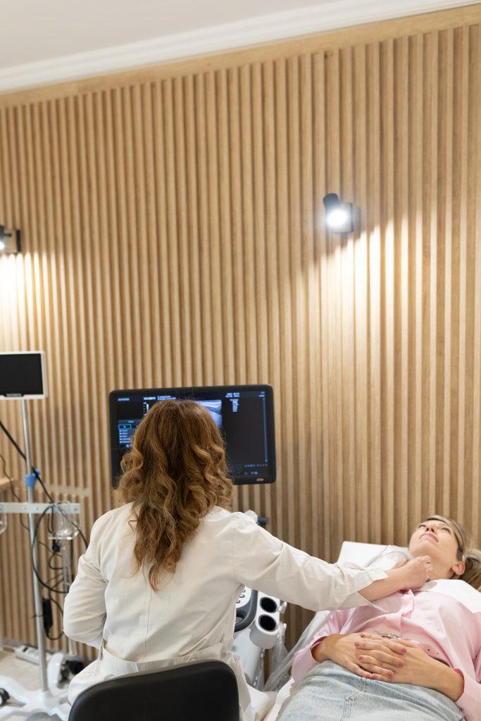
During a head and neck ultrasound, a gel is applied to the skin in the area being examined. This gel helps the ultrasound probe (transducer) make better contact with the skin and ensures the sound waves travel smoothly. The transducer emits high-frequency sound waves that pass through the tissues. These sound waves bounce off the internal structures, and the returning echoes are captured by the transducer. The collected data is then converted into real-time images, which are displayed on a monitor. These images allow the doctor to visualize the tissues and organs in the head and neck area, such as the thyroid gland, lymph nodes, salivary glands, and soft tissues, helping to detect abnormalities like tumors, cysts, or inflammation. The procedure is completely painless and generally takes between 15 to 30 minutes, depending on the area being examined. Since it is non-invasive, there are no risks or side effects associated with the test.
Indications for Head and Neck Ultrasound
Head and Neck Ultrasound is recommended in cases where there are symptoms or indications related to these areas, such as
- Palpable Nodules or Enlarged Lymph Nodes in the Neck or Around the Jaw
- Palpation of Swollen Salivary Glands
- Thyroid Gland Disorders or Swelling
- Neck Pain or Inflammation Unexplained by Other Causes
- Monitoring Known Tumors or Abnormalities in the Head and Neck Area
Areas Examined with Head and Neck Ultrasound:
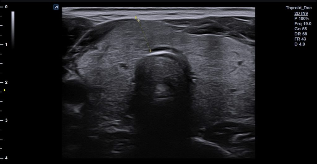
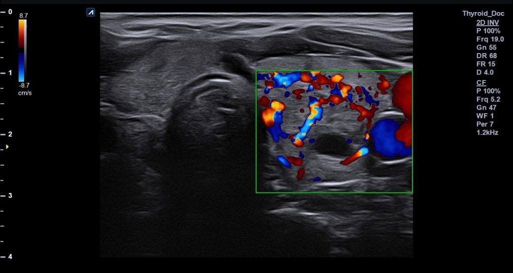
- Thyroid Gland: Ultrasound is used for the detection and evaluation of nodules in the thyroid gland. It helps differentiate between benign and suspicious nodules that may indicate malignancy. Additionally, it is useful for monitoring the progression of both nodules and other thyroid conditions, such as thyroiditis. The evaluation of the gland's structure is also important in cases of hyperthyroidism (overactive thyroid) or hypothyroidism (underactive thyroid). Finally, ultrasound offers the ability for guided biopsy and other interventions, such as fine needle aspiration (FNA) to collect cytological material from suspicious nodules.
- Lymph Nodes: Ultrasound examination of the cervical lymph nodes is an important, non-invasive, painless, and quick diagnostic tool that provides valuable information for the diagnosis and monitoring of lymphatic system disorders. Some situations where ultrasound may be required include swollen lymph nodes, persistent pain or discomfort in the neck, monitoring benign or malignant tumors, detecting infections, and evaluating patients with a history of malignancy.
- Salivary Glands: Ultrasound is useful for detecting inflammations, stones, or tumors in the salivary glands (parotid, submandibular, and sublingual glands). It helps to assess the size, structure, and condition of these glands, aiding in the diagnosis of conditions such as sialolithiasis (salivary stones), sialadenitis (inflammation), and benign or malignant tumors.
- Muscles, Blood Vessels, and Other Tissues of the Neck:The examination allows for the monitoring of soft tissues and blood circulation in the neck area, as well as the identification of potential damage or abnormalities. It can help detect conditions such as muscle tears, vascular issues (e.g., stenosis or aneurysms), and other soft tissue pathologies, providing crucial information for diagnosis and treatment planning.